Mitochondria at a glance
What they are: structures inside most of your cells that turn food and oxygen into usable energy
Where they came from: bacteria that took up residence inside another cell around two billion years ago
Their own DNA: a small genome of 16,569 DNA letters, inherited from your mother
More than power: they also send signals, trigger cell death and alert the immune system
Adaptable: endurance exercise prompts muscle cells to build more of them
Mitochondria are usually introduced as the powerhouses of the cell, and that description is accurate as far as it goes. They produce most of the energy your cells use. But research over the past few decades has shown that they do far more: they carry their own genes, act as signalling hubs, decide when damaged cells should die, warn the immune system of trouble, and constantly reshape themselves in response to demand.
This guide explains what mitochondria are, where they came from, how they make energy, why they carry their own DNA, how cells keep them healthy, how exercise builds more of them, and what research on ageing and NAD has and has not shown. Every key point is linked to its source.
In this guide: What mitochondria are · An ancient partnership · How mitochondria make energy · Their own DNA · More than power plants · Keeping mitochondria healthy · How exercise builds more · Mitochondria and ageing · What about NAD? · What we still do not know · Common questions · References
What mitochondria are
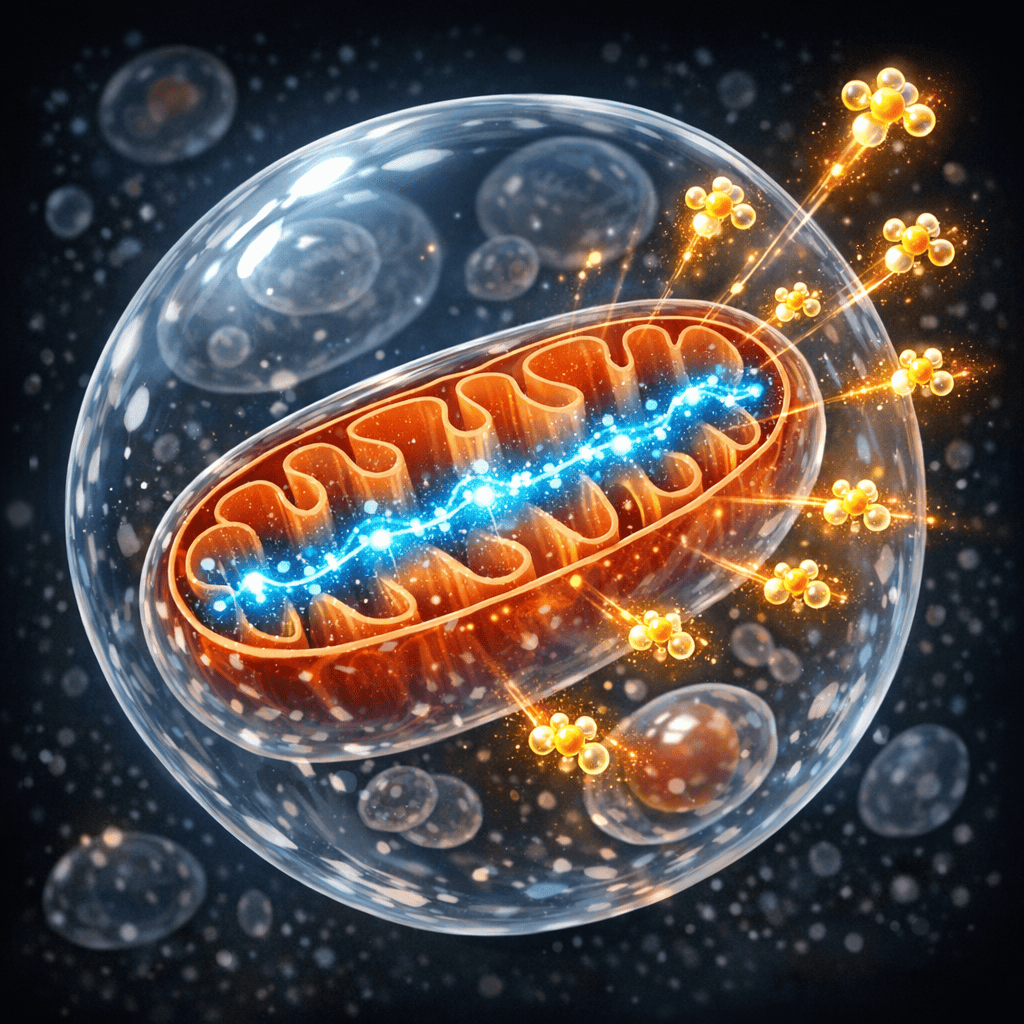
Mitochondria are small structures, or organelles, found in almost every cell in the body apart from mature red blood cells. A single cell can contain anywhere from a few to several thousand, depending on how much energy it needs. Heart muscle, skeletal muscle, liver and brain cells are especially rich in them.
Each mitochondrion has two membranes. The inner membrane is folded into ridges called cristae, which greatly increase its surface area. That inner membrane is where most of the energy production takes place.
An ancient partnership
In 1967, the American biologist Lynn Margulis, then publishing as Lynn Sagan, proposed in the Journal of Theoretical Biology that mitochondria were once free-living bacteria that came to live inside another cell [1]. The idea was controversial at the time but is now widely accepted. It explains why mitochondria have their own DNA, their own double membrane and a way of dividing that resembles bacteria.
That partnership, thought to have begun around two billion years ago, gave complex cells access to a far richer energy supply and is seen by many biologists as a key step in the evolution of animals and plants.
How mitochondria make energy
Mitochondria break down fuel from food and pass the resulting electrons along a chain of protein complexes in the inner membrane, known as the electron transport chain. As the electrons flow, the chain pumps protons across the membrane, building up a difference in charge and concentration, much like water behind a dam. The protons then flow back through an enzyme called ATP synthase, which uses that flow to make ATP, the molecule cells use as their energy currency.
This mechanism was proposed by the British biochemist Peter Mitchell in 1961 in Nature [2]. Called the chemiosmotic theory, it was initially resisted and later earned Mitchell the 1978 Nobel Prize in Chemistry.
The process is efficient but not perfect. A small fraction of electrons leak out of the chain and react with oxygen to form superoxide, a reactive oxygen species. A 2009 review in the Biochemical Journal explains that this leakage depends heavily on conditions inside the mitochondrion and is generally lower when mitochondria are actively making ATP [3]. Some of that reactive oxygen acts as a useful signal; too much contributes to damage, as described in our guide to oxidative stress.
Their own DNA
Mitochondria carry a small circular genome of their own, separate from the DNA in the cell’s nucleus. Its complete sequence was published in Nature in 1981: 16,569 DNA letters encoding two ribosomal RNAs, 22 transfer RNAs and 13 proteins, all of them parts of the energy-producing machinery [4]. The genome is remarkably compact, with almost no space between genes [4]. The great majority of the proteins mitochondria need, however, are encoded in the nucleus and imported.
Mitochondrial DNA is inherited almost entirely from the mother, a finding confirmed in human families in a 1980 study in the Proceedings of the National Academy of Sciences [5]. Because each cell contains many copies, a mutation may be present in some copies and not others, a mixture known as heteroplasmy.
In 1988, Douglas Wallace and colleagues reported in Science the first mitochondrial DNA mutation linked to a human disease, Leber’s hereditary optic neuropathy, a cause of vision loss [6]. Many more have since been identified. A 2015 study in the Annals of Neurology estimated that mitochondrial diseases, caused by mutations in either mitochondrial or nuclear genes, affect at least 1 in 4,300 adults, making them among the most common inherited neurological disorders [7].
In 2025, researchers in Newcastle reported in the New England Journal of Medicine that eight babies had been born after mitochondrial donation, a technique that transfers the parents’ nuclear DNA into a donated egg with healthy mitochondria. Levels of the mother’s harmful mitochondrial variant were undetectable or low in the infants [8]. Long-term follow-up of the children is continuing.
More than power plants
Deciding when cells die. In 1996, a team reported in Cell that cytochrome c, a protein that normally works in the mitochondrial energy chain, is released into the cell when it is under severe stress and helps trigger programmed cell death, or apoptosis [9]. This finding established mitochondria as central decision makers in whether a damaged cell lives or dies.
Alerting the immune system. Because mitochondria descend from bacteria, their DNA resembles bacterial DNA. A 2015 study in Nature showed that when mitochondrial DNA is stressed and escapes into the cell’s interior, it is detected by a sensor called cGAS, which switches on antiviral immune signalling [10]. This is one way damaged mitochondria can contribute to inflammation.
Sending signals. Through reactive oxygen, metabolic by-products and other molecules, mitochondria report on the cell’s energy state to the nucleus and help adjust which genes are switched on [3].
Keeping mitochondria healthy
Mitochondria are not fixed structures. They continually fuse together and split apart. A 2012 review in Science explains that fusion lets partly damaged mitochondria share their contents and compensate for one another, while fission creates new mitochondria and helps isolate damaged parts for removal [11].
Damaged mitochondria are cleared by a form of recycling called mitophagy. Two proteins, PINK1 and Parkin, act as a tagging system: PINK1 builds up on the surface of a damaged mitochondrion and recruits Parkin, which marks it for destruction. Mutations in the genes for both proteins cause inherited forms of Parkinson’s disease, as described in a 2015 review in Neuron, strengthening the case that mitochondrial quality control matters for brain health [12].
How exercise builds more
Cells can increase their number of mitochondria when demand rises, a process called mitochondrial biogenesis. In 1967, John Holloszy reported in the Journal of Biological Chemistry that endurance training in rats substantially increased the mitochondrial enzymes and oxygen-using capacity of their muscles [13], one of the founding findings of exercise biology.
A key controller of this process, PGC-1 alpha, was discovered in 1998 in brown fat, where it rises sharply when mice are exposed to cold and switches on mitochondrial genes [14]. It was later shown to play a similar role in muscle.
The effect is seen in people too. A 2017 study in Cell Metabolism compared 12 weeks of different training types in younger and older adults. High-intensity interval training, alone or combined with resistance training, improved muscle mitochondrial function, and in older adults it reversed many age-related differences in mitochondrial proteins [15].
Mitochondria and ageing
Mitochondrial dysfunction is one of the twelve hallmarks of ageing described in our guide to the biology of ageing. A 2016 review in Molecular Cell describes how declining mitochondrial quality and activity are linked to several other features of ageing, including cellular senescence, chronic inflammation and the decline of stem cells, and how the quality control pathways described above may influence lifespan [16].
What remains unclear is how much of this decline is a cause of ageing rather than a consequence of it, and how much can be changed in people.
What about NAD?
NAD is a molecule that carries electrons in the reactions mitochondria use to make energy, and also acts as a signal for a range of enzymes. A 2018 review in Cell Metabolism notes that NAD levels decline with age in many tissues and that restoring them in old or diseased animals can improve health and, in some studies, extend lifespan [17].
Human evidence is much more limited. A 2018 randomised crossover trial in Nature Communications found that six weeks of nicotinamide riboside, a precursor of NAD, was well tolerated in healthy middle-aged and older adults and raised NAD-related measures in the blood. The authors described the findings on physical function as initial and called for further trials [18]. Raising NAD in blood has not been shown to slow ageing or prevent disease in people.
What we still do not know
Scientists still lack simple ways to measure mitochondrial health in living people, particularly in organs such as the brain and heart. Much of what is known about mitochondrial ageing comes from animals and cells.
The links between mitochondria, inflammation and disease are also still being mapped. It is not yet clear which mitochondrial changes drive conditions such as Parkinson’s disease or type 2 diabetes, and which follow from them [12, 16].
Finally, treatments aimed at mitochondria, from NAD precursors to mitochondrial transplantation, remain largely experimental. Beyond mitochondrial donation for inherited disease [8], very few have reached routine use.
Common questions
Why are mitochondria called the powerhouses of the cell?
Because they produce most of the cell’s ATP, the molecule used to power almost every process in the body [2].
Why do mitochondria have their own DNA?
Because they descend from bacteria that lived inside an ancestral cell [1]. Over time most of their genes moved to the nucleus, but 37 remain [4].
Do we inherit mitochondria from both parents?
No. Mitochondrial DNA is inherited almost entirely from the mother [5].
Can you increase your mitochondria?
Muscle cells build more mitochondria in response to endurance and interval training, a process shown in animals since the 1960s and confirmed in human training studies [13, 15].
Do NAD supplements improve mitochondria?
Animal research is promising, and small human trials show NAD precursors can raise NAD-related measures in blood, but benefits for health in people have not been established [17, 18].
Further reading: Ferroptosis explained: how iron drives a distinct form of cell death.
Further reading: Sleep and cellular repair: what happens inside your cells while you sleep.
Further reading: Redox signalling explained: how cells use reactive oxygen as a message.
Further reading: Inflammation explained · Exercise and your cells
References
- Sagan L. (1967). On the origin of mitosing cells. Journal of Theoretical Biology. https://doi.org/10.1016/0022-5193(67)90079-3
- Mitchell P. (1961). Coupling of phosphorylation to electron and hydrogen transfer by a chemi-osmotic type of mechanism. Nature. https://doi.org/10.1038/191144a0
- Murphy MP. (2009). How mitochondria produce reactive oxygen species. Biochemical Journal. https://doi.org/10.1042/BJ20081386
- Anderson S, Bankier AT, Barrell BG, et al. (1981). Sequence and organization of the human mitochondrial genome. Nature. https://doi.org/10.1038/290457a0
- Giles RE, Blanc H, Cann HM, et al. (1980). Maternal inheritance of human mitochondrial DNA. Proceedings of the National Academy of Sciences. https://doi.org/10.1073/pnas.77.11.6715
- Wallace DC, Singh G, Lott MT, et al. (1988). Mitochondrial DNA mutation associated with Leber’s hereditary optic neuropathy. Science. https://doi.org/10.1126/science.3201231
- Gorman GS, Schaefer AM, Ng Y, et al. (2015). Prevalence of nuclear and mitochondrial DNA mutations related to adult mitochondrial disease. Annals of Neurology. https://doi.org/10.1002/ana.24362
- Hyslop LA, Blakely EL, Aushev M, et al. (2025). Mitochondrial donation and preimplantation genetic testing for mtDNA disease. New England Journal of Medicine. https://doi.org/10.1056/NEJMoa2415539
- Liu X, Kim CN, Yang J, et al. (1996). Induction of apoptotic program in cell-free extracts: requirement for dATP and cytochrome c. Cell. https://doi.org/10.1016/S0092-8674(00)80085-9
- West AP, Khoury-Hanold W, Staron M, et al. (2015). Mitochondrial DNA stress primes the antiviral innate immune response. Nature. https://doi.org/10.1038/nature14156
- Youle RJ, van der Bliek AM. (2012). Mitochondrial fission, fusion, and stress. Science. https://doi.org/10.1126/science.1219855
- Pickrell AM, Youle RJ. (2015). The roles of PINK1, parkin, and mitochondrial fidelity in Parkinson’s disease. Neuron. https://doi.org/10.1016/j.neuron.2014.12.007
- Holloszy JO. (1967). Biochemical adaptations in muscle: effects of exercise on mitochondrial oxygen uptake and respiratory enzyme activity in skeletal muscle. Journal of Biological Chemistry. https://doi.org/10.1016/S0021-9258(18)96046-1
- Puigserver P, Wu Z, Park CW, et al. (1998). A cold-inducible coactivator of nuclear receptors linked to adaptive thermogenesis. Cell. https://doi.org/10.1016/S0092-8674(00)81410-5
- Robinson MM, Dasari S, Konopka AR, et al. (2017). Enhanced protein translation underlies improved metabolic and physical adaptations to different exercise training modes in young and old humans. Cell Metabolism. https://doi.org/10.1016/j.cmet.2017.02.009
- Sun N, Youle RJ, Finkel T. (2016). The mitochondrial basis of aging. Molecular Cell. https://doi.org/10.1016/j.molcel.2016.01.028
- Rajman L, Chwalek K, Sinclair DA. (2018). Therapeutic potential of NAD-boosting molecules: the in vivo evidence. Cell Metabolism. https://doi.org/10.1016/j.cmet.2018.02.011
- Martens CR, Denman BA, Mazzo MR, et al. (2018). Chronic nicotinamide riboside supplementation is well-tolerated and elevates NAD+ in healthy middle-aged and older adults. Nature Communications. https://doi.org/10.1038/s41467-018-03421-7
Redox News Today reports on published research for general interest. This guide is not medical advice. Last reviewed October 2026.

Matt Elliott is the editor of Redox News Today, an independent publication covering peer-reviewed research on cellular health, redox signalling, and related biomedical science.